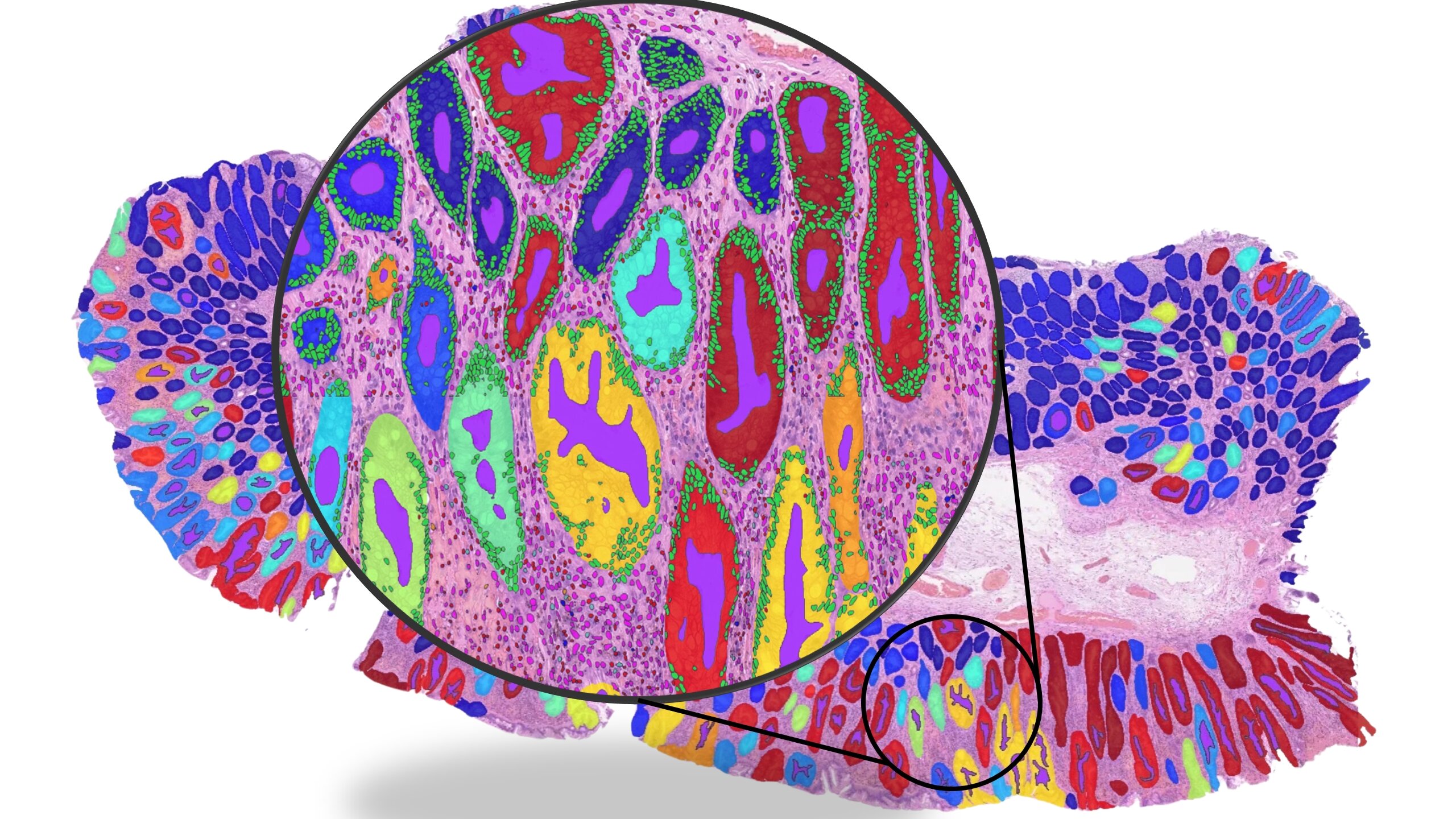
Whole slide image illustrating the detection of key histological structures such as glands and cells.
A recent study from the University of Warwick, published in Nature Biomedical Engineering, evaluates the reliability of artificial intelligence (AI) tools designed to predict cancer biology directly from histopathology images. These tools are often positioned as enabling faster and more cost-effective diagnostics. The study analysed over 8,000 patient samples across multiple cancer types, including breast, colorectal, lung and endometrial cancers. While several AI models demonstrated high predictive accuracy, the findings indicate that this performance is frequently driven by “shortcut learning” where models rely on indirect correlations rather than biologically meaningful signals.
For example, instead of identifying a specific mutation such as BRAF, models may detect associated features like micro-satellite instability and use these as proxies. This approach can produce accurate predictions in controlled datasets but may fail when such correlations do not hold in different clinical settings. The study highlights that current AI pathology tools may therefore lack robustness for routine clinical use. It also underscores the limitations of relying on headline accuracy as a primary performance metric, as this may mask underlying biases and confounding factors in model behaviour.
The authors emphasise the need for more rigorous and bias-aware evaluation frameworks to ensure that AI systems capture genuine biological relationships. Strengthening validation approaches will be essential to support the safe and effective integration of AI into clinical oncology and to ensure that these tools provide meaningful value beyond existing diagnostic methods.
________________________________________________________________________________________________________________
Article can be accessed on: MedicalXpress
Image credit: Dr. Fayyaz Minhas / University of Warwick.