An international consortium led by Howard Hughes Medical Institute investigator Vamsi Mootha has mapped mitochondrial proteins across five groups of disease-causing organisms, uncovering potential drug targets and new insights into mitochondrial evolution.
The Project examined Giardia, Acanthamoeba, trypanosomes, Leishmania and Babesia, alongside a plant. Using protein correlation profiling and mass spectrometry, researchers identified proteins associated with mitochondria across the organisms. The findings were reported in nine scientific papers. The team identified 33 protein families shared by at least two of the parasites but absent from human mitochondria. These represent potential targets for treatments against multiple parasitic diseases. However, the researchers emphasised that identifying targets is an early step and that further work is needed before any medicines can be developed.
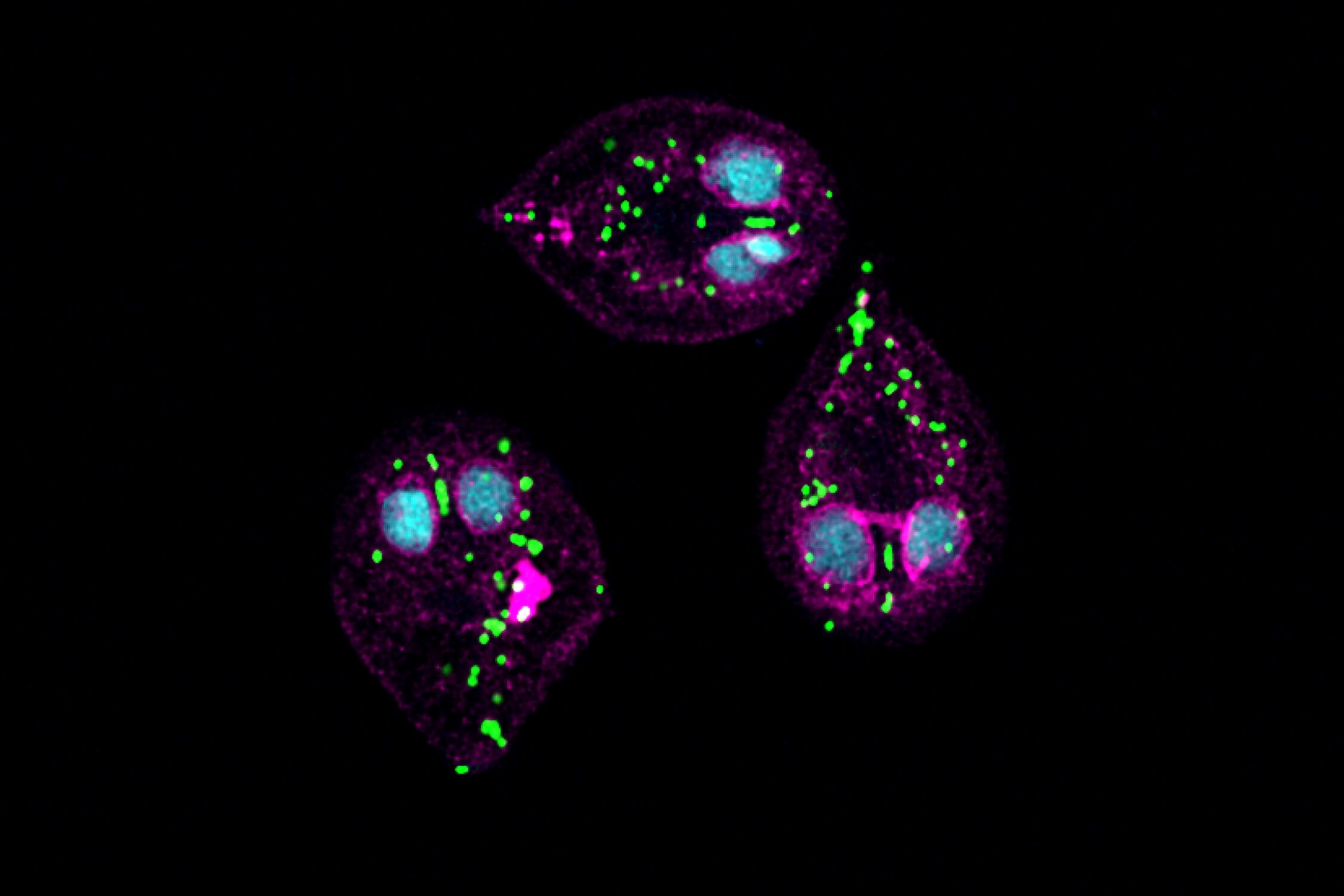
The study also revealed substantial differences in mitochondrial structure and function. Giardia’s simplified mitochondrion-related organelles contain 59 proteins and have no mitochondrial DNA, with many protein functions remaining unknown. In contrast, Acanthamoeba mitochondria can use an enzyme called hydrogenase to produce hydrogen gas when oxygen is scarce. Researchers also trained an artificial intelligence model to predict mitochondrial protein sets in nearly 200 eukaryotes. Their comparative analysis suggested that mitochondrial DNA was lost independently in five evolutionary lineages, accompanied by reductions in organelle size.
The findings further suggest that mitochondria in the last common ancestor of living eukaryotes were metabolically flexible and could generate energy with or without oxygen. The publicly available data provide a valuable resource for broader future investigations into mitochondrial biology and evolution.
_________________________________________________________________________________________________________
The article can be accessed on: MedicalXpress
Image: Giardia lamblia, which causes giardiasis, shown here in its active, dividing form. The green dots mark the simplified mitochondrion-related organelles. Credit: Manish Goyal, Samuelson Lab / Boston University